New microscope reveals living cells in unprecedented detail
On Mar. 2, 2026, Stanford scientists announced that a one-of-a-kind instrument can show cell structures interacting in real time at high resolution without fluorescent labels, potentially enabling breakthroughs in a range of life science fields.
The researchers have combined two microscopy techniques to create a one-of-a-kind instrument that can show cell structures interacting in real time at an unprecedented 120-nanometer resolution – the highest achieved without the use of fluorescent labels.
This new “label-free” technology, called Interferometric Image Scanning Microscopy, or iISM, will allow scientists to observe cellular structures in their wider context, including their responses to intrusions, such as pathogens or drugs. The advance is detailed in the Nature portfolio journal Light: Science and Applications.
“This new microscope provides a fantastic new view into the cell, where you can see the tiny structures and machines in the cell moving, changing, and interacting without having to add fluorescence to observe them,” said senior author W.E. Moerner, the Harry S. Mosher Professor of Chemistry in Stanford’s School of Humanities and Sciences. “It’s a wonderful look into these complex little cellular boxes that drive our life.”
The iISM’s capabilities could enable breakthroughs in a range of life science fields, from understanding disease mechanisms and developing drugs to investigating relationships between plants and microbes.The iISM manages to achieve higher resolution and sensitivity by combining the advantages of two different microscopy methods – a combination that speaks to the expertise of the two co-authors. Moerner, who won the 2014 Nobel Prize in chemistry for his work on super-resolution fluorescence microscopy, specifically sought to bring Kueppers to Stanford because of her doctoral work focused on
“interferometric scattering microscopy.”
They have already started three collaborations with other Stanford researchers. One uses the new microscope to see the interaction among plant cells, fungi, and bacteria in real time. Another collaboration uses the iISM to see how a cancer drug is taken up by a cell, and a third planned effort will investigate how red blood cells change shape when encountering a malaria infection.
Tags:
Source: Stanford University
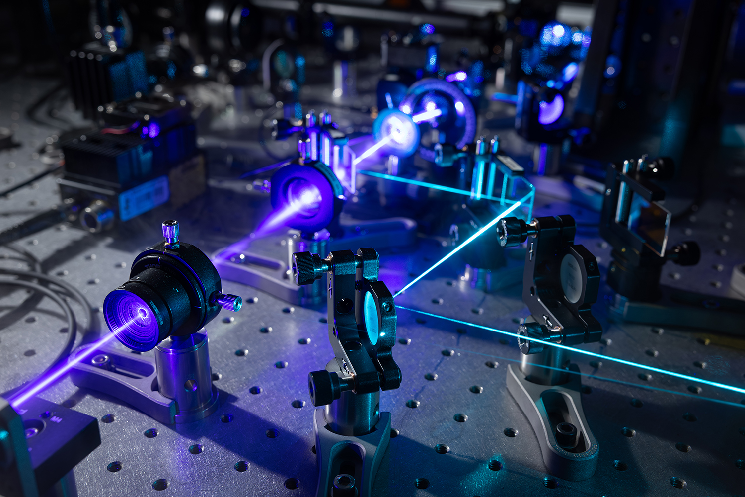
Credit: Photo: Detail of the laser array that is part of the Interferometric Image Scanning Microscopy technology. Courtesy: Jim Gensheimer, Stanford University.