World first – First ever transplant of frozen testicular tissue after chemotherapy during childhood provides hope for fertility restoration
On Jan. 9, 2025, for the first time ever, cryopreserved immature testicular tissue was reintroduced after 16 years in an infertile man who underwent chemotherapy in childhood. With this transplant, doctors and researchers hope to trigger sperm cell production and restore the patient’s fertility. The procedure was successful, and the patient is recovering well. In one year from now, the team will check for the presence of mature sperm. This transplant is the outcome of years of research of Vrije Universiteit Brussel and Brussels IVF, the centre for reproductive medicine of UZ Brussel. It is the consequence of another world first, because our hospital was the first worldwide to launch a cryopreservation programme for testicular tissue in 2002.
In boys who undergo radical treatments before starting puberty that can affect their fertility, testicular tissue can be preventively removed and frozen. This is done to preserve testicular stem cells, the precursors of sperm cells. Chemotherapy and radiation therapy can destroy these cells, leading to infertility later in life.
Prepubescent boys do not yet produce sperm cells. The removed tissue contains stem cells that would normally produce sperm after starting puberty. The cryopreservation of tissue in young boys means we can potentially restore fertility later in life with a transplant.
Researchers of Vrije Universiteit Brussel and Brussels IVF, the centre for reproductive medicine of UZ Brussel, have now reintroduced several tissue fragments in a man who underwent treatment in childhood with chemotherapy which negatively affected his fertility. The procedure is part of a research project, funded by the Research Foundation Flanders (FWO) and the VUB (SRP-groeier), to assess whether such a transplant effectively restores fertility.
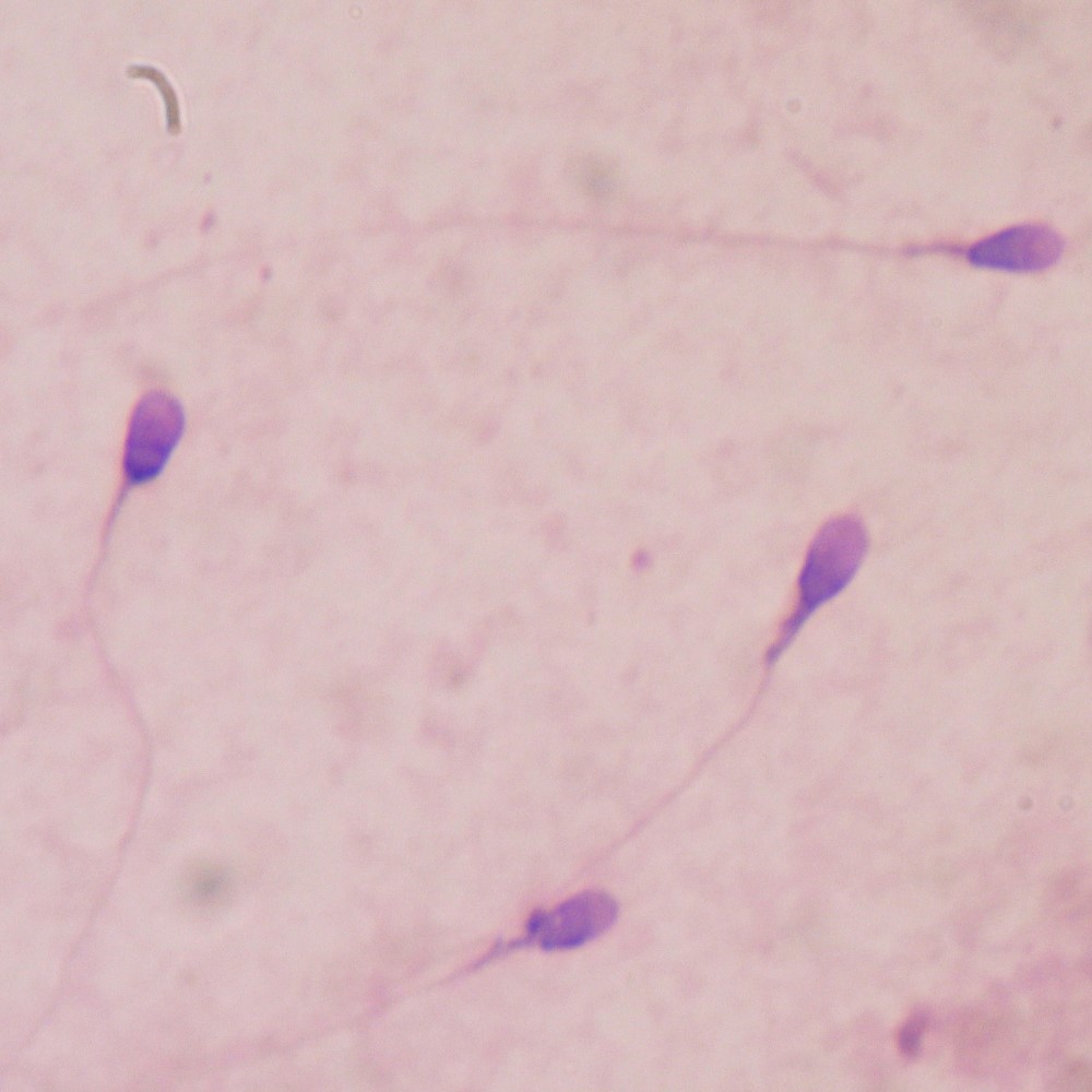
The transplant of testicular tissue consists of the reintroduction of four tissue fragments in the testicle and four in the scrotum. The technique is designed to make the patient’s body produce sperm cells autonomously. After the transplant, the patient will be monitored for one year, by means of blood work, hormone determinations, ultrasound examination and a semen sample every three months. The semen sample is examined for the presence of sperm. Because the tissue fragments are not directly connected to the sperm duct, the researchers do not expect sperm cells to naturally find their way into the semen sample. If the patient wishes to have children, ART will be necessary.
Tags:
Source: Vrije Universiteit Brussel
Credit: