Scientists map how the flu virus rewires the human cell from the inside
On Jul. 20, 2026, researchers at EMBL Hamburg and collaborators at the Leibniz Research Institute for Molecular Pharmacology (FMP) announed they have mapped how the influenza A virus rewires infected human cells in unprecedented detail. To do this, the researchers used a customised experimental workflow to directly observe how proteins interact inside intact infected cells.
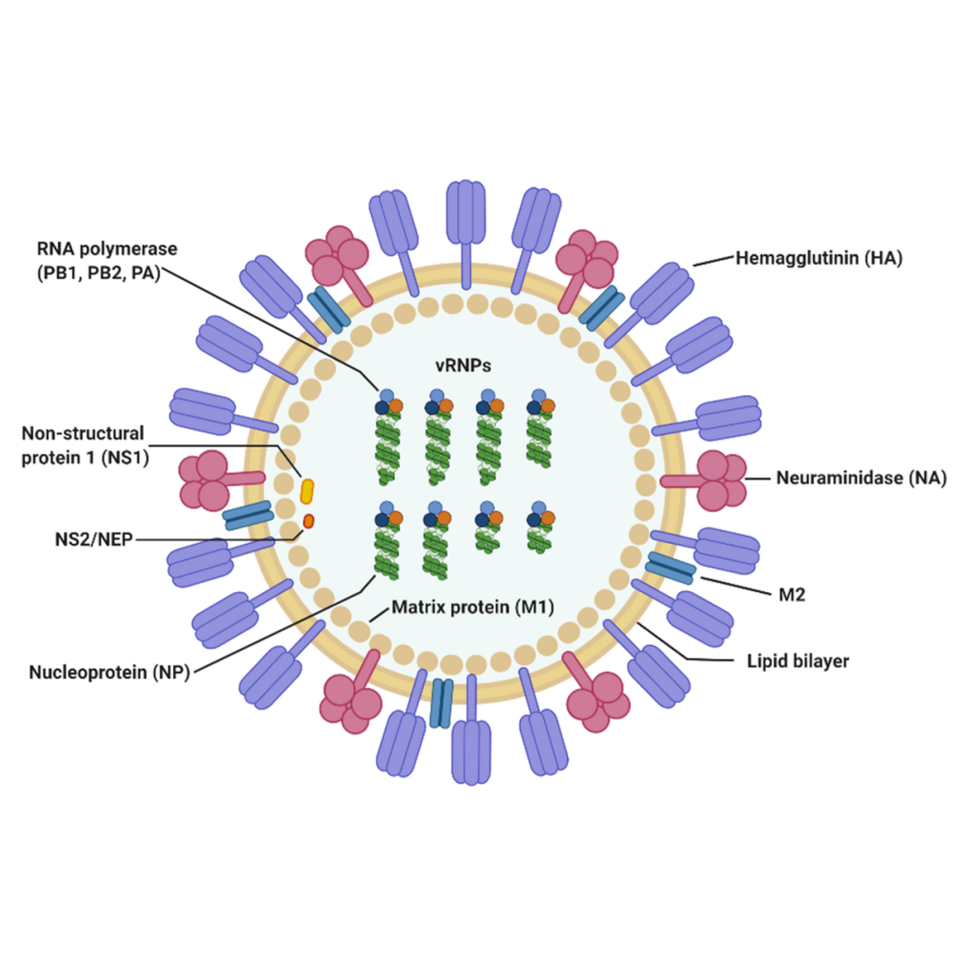
Every year, seasonal influenza kills up to 650,000 people globally and causes serious illness for 3–5 million individuals. The influenza A virus, in particular, has been responsible for several pandemics, including the 1918 Spanish Flu pandemic. When this virus infects cells, it releases its genetic material, called RNA, which contains blueprints for a handful of proteins. These proteins then spread throughout the host cell and repurpose its molecular machinery to make more viruses.
Scientists want to understand this process in detail, as it would help in designing better drug therapies and vaccines against the flu virus. That’s why it’s crucial to figure out how proteins of the flu virus interact with proteins of host cells and subvert them to meet the virus’s needs. This is the first time that scientists have mapped direct virus-host protein contacts at scale inside intact influenza-infected cells, with enough structural detail to model how the proteins fit together.
Studying protein-protein interactions in action during infection is easier said than done. Most previous studies relied on biochemical methods that shared one limitation: the cell had to be broken open before the interactions could be measured. Once the cell’s compartments were gone, proteins that were never in contact inside the cell could meet in the test tube, and fragile or location-specific contacts could be lost. It was then hard to know which interactions actually happened inside an infected cell.
“This is when we learned that our collaborators – Boris Bogdanow and Fan Liu – at FMP Berlin had developed a specialised version of cross-linking mass spectrometry (XL-MS), a long-established technique for mapping protein contacts, tailored specifically to virus-infected cells,” said Kosinski. This was the critical breakthrough. It allowed researchers to do what previous methods couldn’t, including capturing short-lived and location-specific interactions.
“XL-MS allows us to capture protein-protein interactions directly in infected intact cells, while also providing structural information about how these interactions are happening,” explained Bogdanow, who is now a Junior Research Group Leader at the Institute of Virology, Charité – Universitätsmedizin Berlin. “This gives us insight into the interface between the virus and the human cell and may, through structural modelling, help identify actionable targets for future pharmaceutical interventions.”
By combining the results obtained through XL-MS with computational structural modelling, the researchers could not only identify which viral and human proteins interact, but also predict how they physically fit together. For this, they used a modified version of AlphaFold, the Nobel prize-winning protein structure prediction algorithm.
Published in Nature Microbiology, the study results uncovered two important ways in which the virus hijacks the cell. The first one involves haemagglutinin, a protein on the virus’s surface that it uses to bind and enter host cells. The researchers traced how haemagglutinin moves through the cell’s internal transport and processing system. This is a network of compartments that modifies and prepares proteins before they are shipped to their final destination. This revealed how host proteins, some with previously unknown functions, helped the virus correctly fold and modify haemagglutinin during infection.
The second one involves paraspeckles, small droplet-like compartments in the nucleus. The researchers found that infection by the influenza A virus causes these organelles to dissolve, releasing the RNA-binding proteins bound within them, which the virus can then use to replicate.
Tags:
Source: EMBL
Credit: