Atlas of the brain’s striatum could guide researchers to new drug treatments
On Sept. 1, 2026, Massachusetts Institute of Technology (MIT) researchers release a study that reveals insights into populations of neurons affected by Huntington’s disease, schizophrenia, addiction, and other disorders.
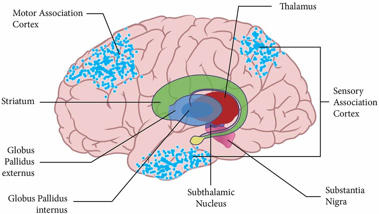
A region of the brain called the striatum is critical for many cognitive and motor functions, including decision-making, control of movement, habit formation, and processing of reward. It also plays a role in addiction and is significantly affected by Huntington’s disease, schizophrenia, and other disorders.
In work that could help scientists devise new treatments for those diseases, MIT researchers have generated a new atlas of the neurons found within the striatum. Using single-cell RNA sequencing and other techniques, they were able to identify 31 subgroups of neurons based on which genes they express.
These groups include neurons that are involved in addiction, depression, and schizophrenia. The researchers also discovered why some neurons of the striatum are more vulnerable to Huntington’s disease. All of these results, the researchers say, could help scientists develop new drugs to combat these conditions.
Another key finding of the paper helps to shed light on why the dorsal (upper) part of the striatum is more vulnerable to Huntington’s disease. The disease is caused by an inherited version of the huntingtin gene that carries too many repetitive DNA segments, called CAG repeats.
The researchers found that dorsal populations of medium spiny neurons express higher levels of the genes MSH2 and MSH3, which play a role in increasing the number of CAG repeats found in the huntingtin gene. As more of those repeats accumulate, the mutated version of the huntingtin protein becomes more harmful to cells.
The researchers hope that this map, built from tissue contributions by brain donors and their families, and assembled across disciplines and institutions, will provide an important starting point for researchers pursuing new treatments for some of the most difficult-to-treat brain disorders.
Tags:
Source: Massachusetts Institute of Technology
Credit: Illustration: Cortical surface with an overlay of the basal ganglia and thalamus, Courtesy: Colder B., 2015, Wikipedia..