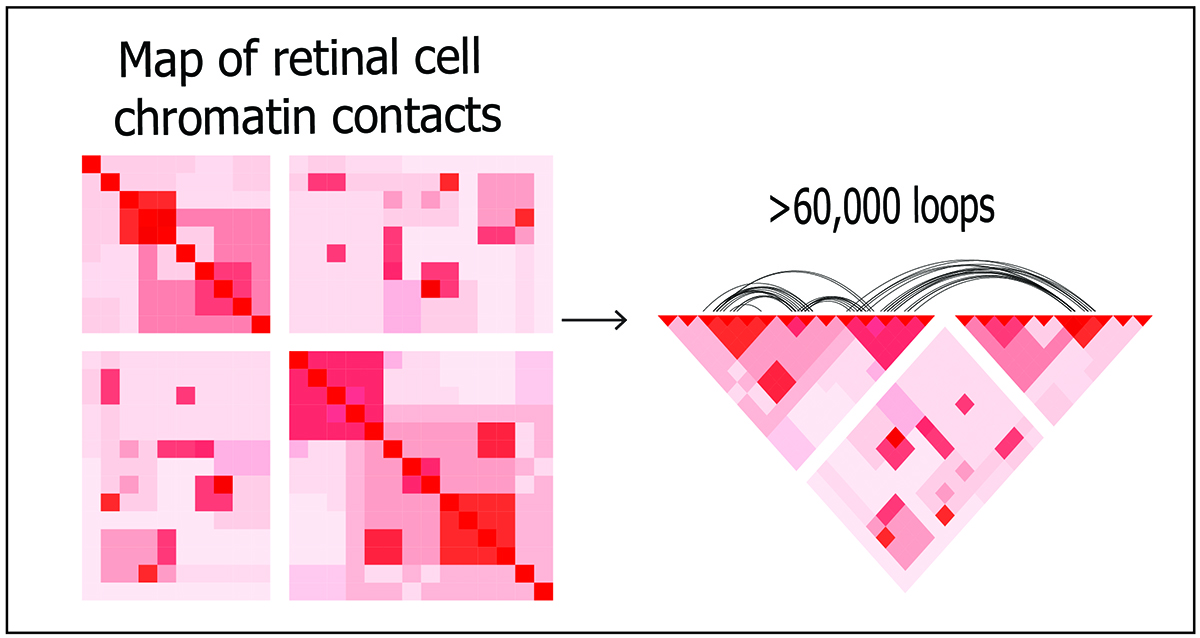
3D map revealed DNA organization within human retina cells
On Oct. 7, 2022, the National Eye Institute researchers announced they had mapped the organization of human retinal cell chromatin, the fibers that package 3 billion nucleotide-long DNA molecules into compact structures that fit into chromosomes within each cell’s nucleus. The resulting comprehensive gene regulatory network provides insights into regulation of gene expression in general, and in retinal function, in both rare and common eye diseases. The study published in Nature Communications.
Adult human retinal cells are highly specialized sensory neurons that do not divide, and are therefore relatively stable for exploring how the chromatin’s three-dimensional structure contributes to the expression of genetic information.
Chromatin fibers package long strands of DNA, which are spooled around histone proteins and then repeatedly looped to form highly compact structures. All those loops create multiple contact points where genetic sequences that code for proteins interact with gene regulatory sequences, such as super enhancers, promoters, and transcription factors.
Such non-coding sequences were long considered “junk DNA.” But more advanced studies demonstrate ways these sequences control which genes get transcribed and when, shedding light on the specific mechanisms by which non-coding regulatory elements exert control even when their location on a DNA strand is remote from the genes they regulate.
Using deep Hi-C sequencing, a tool used for studying 3D genome organization, the researchers created a high-resolution map that included 704 million contact points within retinal cell chromatin. Maps were constructed using post-mortem retinal samples from four human donors.
The researchers then integrated that chromatin topology map with datasets on retinal genes and regulatory elements. What emerged was a dynamic picture of interactions within chromatin over time, including gene activity hot spots and areas with varying degrees of insulation from other regions of DNA.
They found distinct patterns of interaction at retinal genes suggesting how chromatin’s 3D organization plays an important role in tissue-specific gene regulation.
The researchers integrated the chromatin topology map with data on genetic variants identified from genome-wide association studies for their involvement in AMD and glaucoma, two leading causes of vision loss and blindness. The findings point to specific candidate causal genes involved in those diseases.
The integrated genome regulatory map will also assist in evaluating genes associated with other common retina-associated diseases such as diabetic retinopathy, determining missing heritability and understanding genotype-phenotype correlations in inherited retinal and macular diseases.
Tags:
Source: National Institutes of Health
Credit: