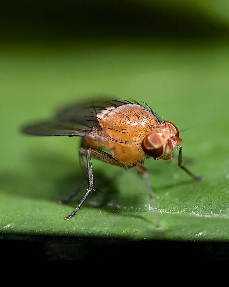
Complete fly brain imaged at nanoscale resolution
On Jul. 19, 2018, scientists at Howard Hughes Medical Institute (HHMI) announced detailed imaging of the entire brain of an adult female fruit fly using transmission electron microscopy. Researchers can now trace the path of any one neuron to any other neuron throughout the whole brain, says neuroscientist Davi Bock, a group leader at Janelia who reported the work along with his colleagues in the journal Cell.
The Janelia team’s data offers a new tool for scientists racing to map these connections. And, in a memory center of the brain, the data also revealed a new cell type and other surprises. “Any time you look at images with higher resolution and more completeness, you’re going to discover new things,” Bock says.
The fruit fly brain, roughly the size of a poppy seed, contains about 100,000 neurons (humans have 100 billion). Each neuron branches into a starburst of fine wires that touch the wires of other neurons. Neurons talk to one another through these touchpoints, or synapses, forming a dense mesh of communication circuits.
Scientists can view these wires and synapses with an imaging technique called serial section transmission electron microscopy. First, they infuse the fly’s brain with a cocktail of heavy metals. These metals pack into cell membranes and synapses, ultimately marking the outlines of each neuron and its connections. Then the researchers hit slices of the brain with a beam of electrons, which passes through everything except the metal-loaded parts. “It’s the same way that x-rays go through your body except where they hit bone,” Bock explains.
He and a crew of scientists developed new tools to speed up the process. The team used high-speed cameras and two custom-built systems to rapidly move tissue samples in eight-micrometer increments, allowing them to quickly capture images of neighboring areas. They were able to image an entire brain slice in less than seven minutes – five times faster than the previous high-throughput transmission electron microscope camera array, TEMCA1. Bock and his colleagues also benefitted from a custom robotic loader built at Janelia that picks up and places samples automatically.
The millions of images Bock’s team collected and stitched together offer an in-depth look at the fly brain – and the chance to explore uncharted areas. Bock’s team traced the paths of neurons that reach out to the mushroom body, a region involved in memory and learning. These cells, called olfactory projection neurons, have been well described previously, using light microscopy. Manually tracing the outlines of these neurons and all their wirelike projections let Bock’s team confirm the quality of their image data.
A better understanding of this brain circuitry could give scientists insight into fly behavior, Bock says. “We think it will tell us something about how the animal learns – how it associates odors with a reward or punishment,” he explains. Now, more than 20 lab groups are digging into the new dataset, tracing neurons and outlining the brain’s circuitry. Bock calls the data a resource that’s free to be mined by neuroscientists probing the mind of the fly. “It adds another tool to the toolkit we’re using to understand this animal.”
Tags:
Source: Howard Hughes Medical Institute
Credit: