First human endothelial cell atlas maps how blood vessels age
On Oct. 27, 2026, to uncover how endothelial cells, the inner linings of blood vessels. change over the course of a lifespan, an international research team led by Laura de Rooij at the CeMM Research Center for Molecular Medicine of the Austrian Academy of Sciences announced they have created the Human Aging Endothelial Cell Atlas, the first comprehensive atlas of vascular aging. By analyzing gene activity data from more than 375,000 human cells across 290 samples and 12 tissues, the scientists reveal that aging affects different parts of the vascular system in distinct ways. The study, published in Nature Aging, provides new insights into the biological processes underlying vascular aging and a valuable resource for future research into age-related diseases and vascular repair.
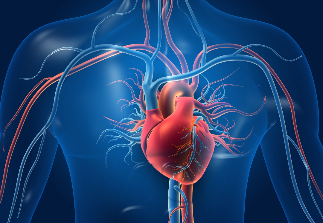
With every heartbeat, blood is pumped through a vast network of vessels throughout our body, delivering vital oxygen, nutrients, and other essential substances to even its most remote corners. But these vessels are far more than passive tubes: Their inner walls are covered by a thin layer of specialized cells called endothelial cells, which regulate blood flow, control what passes into surrounding tissues and help coordinate responses to injury. They even contribute to the specific functions of the organs they serve, such as forming a protective barrier in the brain or supporting filtration in the kidneys. Yet, as we grow older, this finely tuned system can become dysfunctional, increasing the susceptibility to cardiovascular disease and its associated mortality.
Although endothelial cells are essential for vascular and organ function, they are often overlooked in studies of aging. As such, what happens inside endothelial cells as we age, and why their function may deteriorate over time, remains poorly understood. This is the question CeMM Principal Investigator Laura de Rooij and her team set out to answer. Rather than studying blood vessels as a whole, they examined the activity of genes in individual endothelial cells. This approach, known as single-cell analysis, makes it possible to see which genes are active in each cell, and how their activity changes with age. By bringing together data from previously published single-cell studies, the team saw an opportunity to take a closer look at these cells and uncover patterns of vascular aging that might otherwise have remained hidden.
In their latest study, published in Nature Aging and led by first author Sarah Dobner, a PhD student in de Rooij’s group, the team brought together data from 34 existing studies, covering more than 375,000 cells from 290 samples across 12 human tissues. By focusing on tissues classified as healthy or non-diseased, they aimed to identify molecular changes associated with aging rather than changes primarily linked to diagnosed disease. The result is the Human Aging Endothelial Cell Atlas, a resource that allows scientists to examine how different populations of vascular cells change across the human body.
A particularly intriguing observation concerned the cells lining veins. These cells can give rise to sprouts that develop into new blood vessels, an important part of vascular renewal. Yet in older individuals, they showed reduced activity of several genes involved in vessel formation. This could help explain why blood vessels may become less capable of renewing themselves with age, although the study did not directly measure repair capacity. Across multiple tissues, the researchers also observed increased activity of genes associated with inflammation and cellular senescence, a state in which cells stop dividing and can influence their surroundings.
Tags:
Source: News-Medical.net
Credit: