USC study reveals differences in early Alzheimer’s brain markers across diverse populations
On Mar. 12, 2026, a team of researchers at the University of Southern California Mark and Mary Stevens Neuroimaging and Informatics Institute (Stevens INI) at the Keck School of Medicine has identified important differences in how early Alzheimer’s disease-related brain changes appear across racial and ethnic groups, underscoring the need for more inclusive approaches to studying and diagnosing the disease. Their findings are now available in Alzheimer’s & Dementia: The Journal of the Alzheimer’s Association.
In a large, racially and ethnically diverse study of older adults without dementia, researchers found that Black and Hispanic participants showed higher levels of tau, a protein linked to Alzheimer’s, in key memory-related regions of the brain compared to non-Hispanic white participants, even before the buildup of amyloid plaques typically associated with Alzheimer’s disease. However, the relationship between these brain proteins and memory performance varied across groups, suggesting that Alzheimer’s biomarkers may not tell the full story for everyone.
The findings come from the Health and Aging Brain Study–Health Disparities (HABS-HD), one of the largest and most diverse brain-imaging studies of aging in the US and were made possible by advanced PET brain scans that can detect abnormal protein buildup years before symptoms appear.
“Most Alzheimer’s research has been based on non-Hispanic white participants, and our results show that we can’t assume those patterns apply equally to everyone,” said Koral V. Wheeler, MS, lead author of the study and PhD candidate at the Stevens INI. “If we want to advance precision medicine efforts for all communities, we need to understand how these brain markers behave across diverse populations.”
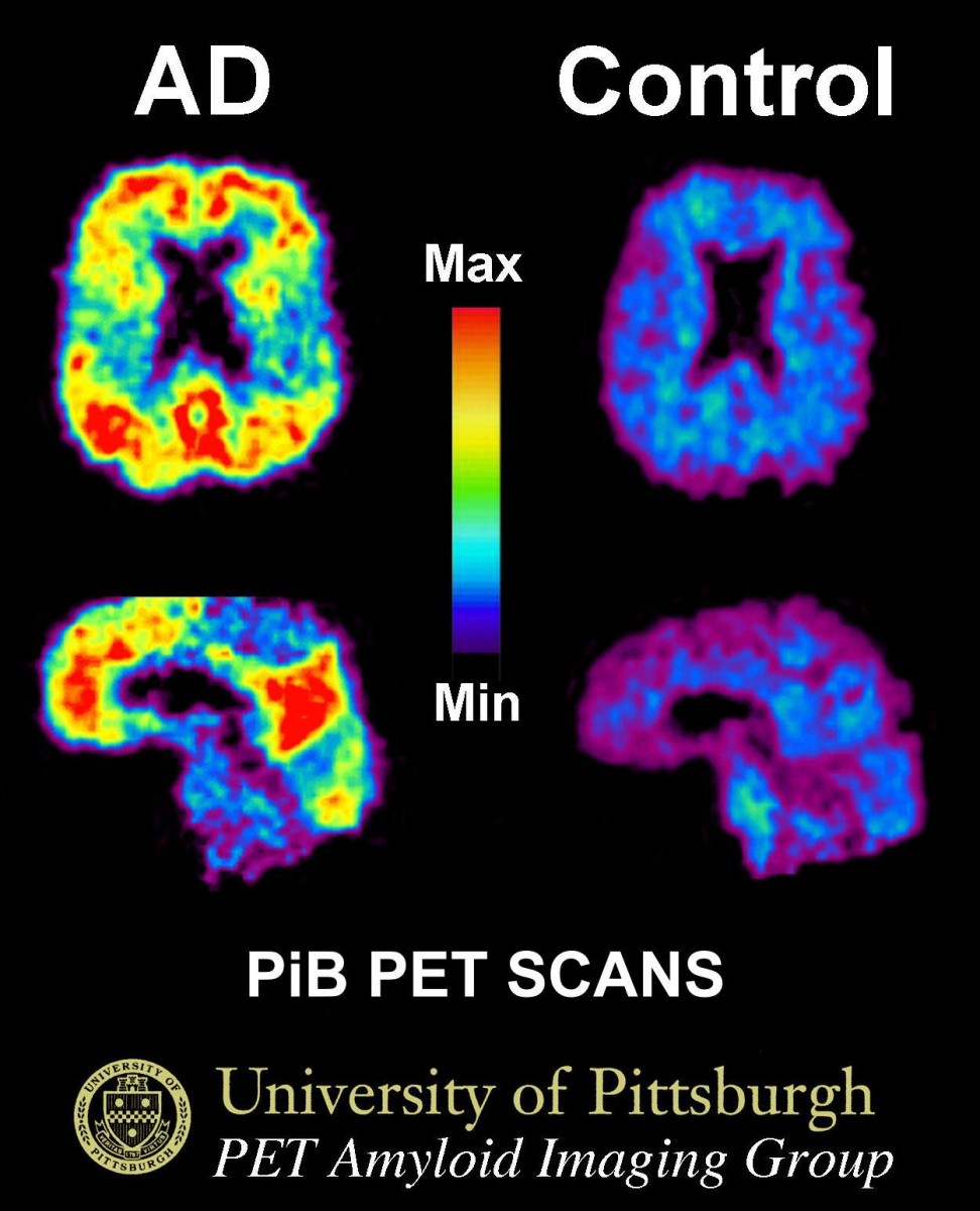
Alzheimer’s disease is characterized by the buildup of two proteins in the brain: amyloid beta, which forms plaques, and tau, which forms tangles that disrupt communication among brain cells. Tau accumulation in the medial temporal lobe, a region critical for memory, is considered an early warning sign of disease progression.
Using a newer generation tau PET tracer, the research team examined brain scans and memory test results from more than 1,500 adults who were cognitively normal or had mild cognitive impairment. A tau PET tracer is a specialized radioactive imaging agent used in PET scans to detect and visualize the accumulation of abnormal tau protein tangles in the brain. While higher tau levels were linked to worse memory overall, amyloid buildup strengthened this link only in non-Hispanic white and Hispanic participants, not in Black participants.
The study also found that some of the observed differences in tau levels may have reflected limitations in the scan itself, rather than true biological differences. In some cases, the tracer can produce signals in nearby areas that are not actually related to tau, making the images harder to interpret. This highlights the importance of carefully validating imaging tools in diverse populations.
These results point to different biological pathways and risk profiles for developing cognitive decline that may shape how the disease develops in different populations and which would affect how it should be detected and treated.
Future studies will follow participants over time to better understand how tau, amyloid, vascular health, genetics, and social determinants interact to influence cognitive aging across communities.
Tags:
Source: Keck School of Medicine of USC
Credit: Image: PiB-PET scan of a patient with Alzheimer’s disease on the left and an elderly person with normal memory on the right. Areas of red and yellow show high concentrations of PiB in the brain and suggest high amounts of amyloid deposits in these areas. Courtesy: University of Pittsburgh.