Scientists develop a new method to study gene function in cells and tissue
On Oct. 7, 2024, Columbia University researchers announced they had developed a new optical pooled screening approach called CRISPRmap, which enables the coupling of optical properties of single cells to targeted genetic perturbations.
Optical phenotypes are typically inaccessible for sequencing-based approaches based on cell lysis but include crucial information such as cell morphology, protein subcellular localization, cell-cell interactions, extracellular matrix factors, and tissue organization.
CRISPRmap allows for spatially resolved interrogation of gene function in tissues, enabling researchers to map both cell-intrinsic and cell-extrinsic effects of perturbations, which are not accessible through in vitro studies. The study findings were published in Nature Biotechnology.
Tags:
Source:
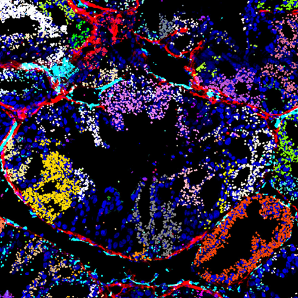
Credit: Photo: Mapping Multimodal Phenotypes to Perturbations in Cells and Tissue with CRISPRmap. Visualization of in vivo CRISPRmap barcode detection on a xenograft tumor section, showing the guide distribution in the tumor microenvironment. Decoded CRISPR guide-identifying barcodes detected by CRISPRmap are shown as spots false-colored according to their guide identity. Courtesy: Gaublomme Lab, Columbia University.