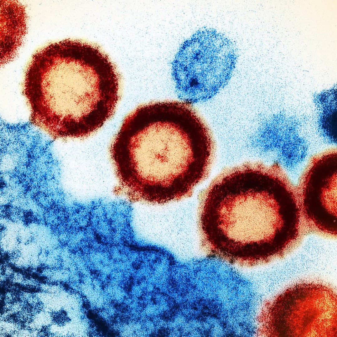
Electron microscopy technique revealed how a key human antibody disarms many strains of HIV
On Apr. 29, 2020, Scripps researchers announced they had developed a method for examining the machinery of HIV in unprecedented detail; and in a near-native environment that hasn’t been possible before. They were able to see for the first time the complete picture how a well-known human antibody attacks a vulnerable site protruding from HIV’s sleek membrane surface.The study, which was published in Cell Reports, looks specifically at HIVメs fusion proteins; these spiky proteins are in charge of initiating infection of healthy immune cells in a patient’s body.
In 2-D images and 3-D structures captured with Scripps Research’s electron microscopes, the team saw precisely how the antibody, known as 10E8, attacks HIV’s fusion protein. The microscopy showed that the antibody wedges itself into the protein’s base (an area officially known as the “membrane proximal external region,” or MPER) and bends the spike before lifting it from the virus’s lipid surface to render it ineffective, Rantalainen explains.
The vulnerable MPER of the fusion protein is conserved across most strains of HIV. This is important because HIV mutates rapidly and escapes the antibody response. For a therapeutic antibody to be effective, it must attack a region that’s consistent across all or most strains. For that reason, this class of antibodies is of special interest to vaccine developers.
The new structural biology study is significant not only for the knowledge it provides to scientists looking to develop a vaccine for HIV, Rantalainen says, but because it presents a valuable new technique for using electron microscopy to observe fusion proteins in a more natural membrane environment.
Previously, structural studies of HIV spike proteins have been hindered by the difficulties in imaging the full protein as it protrudes from the virus’ waxy, double-layered outer membrane. Cryo-EM imaging works by flash-freezing proteins and then fixing them in films of glassy ice, but production of full spike samples for electron microscopy in a membrane has not been successful so far.
Tags:
Source: Scripps Research
Credit: