New hybrid custom-designed Microscope Can Image Full 3D Orientation & Position of Molecules in Cells
On Feb. 21, 2025, a team of researchers from the Marine Biological Laboratory (MBL) announced a hybrid custom-designed diSPIM microscope that for the first time allows scientists to simultaneously image the full 3D orientation and position of an ensemble of molecules, such as labeled proteins inside cells.
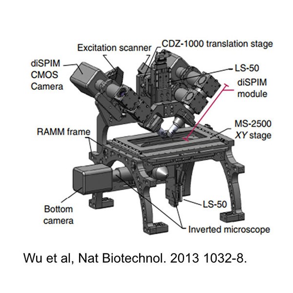
The microscope combines polarized fluorescence technology, a valuable tool for measuring the orientation of molecules, with a dual-view light sheet microscope (diSPIM), which excels at imaging along the depth (axial) axis of a sample.
This scope can have powerful applications. For example, proteins change their 3D orientation, typically in response to their environment, which allows them to interact with other molecules to carry out their functions.
The team hopes to make their system faster so that they can observe how the position and orientation of structures in live samples change over time. They also hope development of future fluorescent probes will enable researchers to use their system to image a greater variety of biological structures.
The research was published in the Proceedings of the National Academy of Sciences.
Tags:
Source: University of Chicago, Marine Biological Laboratory
Credit: