University of Pittsburgh researchers Reveal most detailed images ever of bacteria-killing viruses
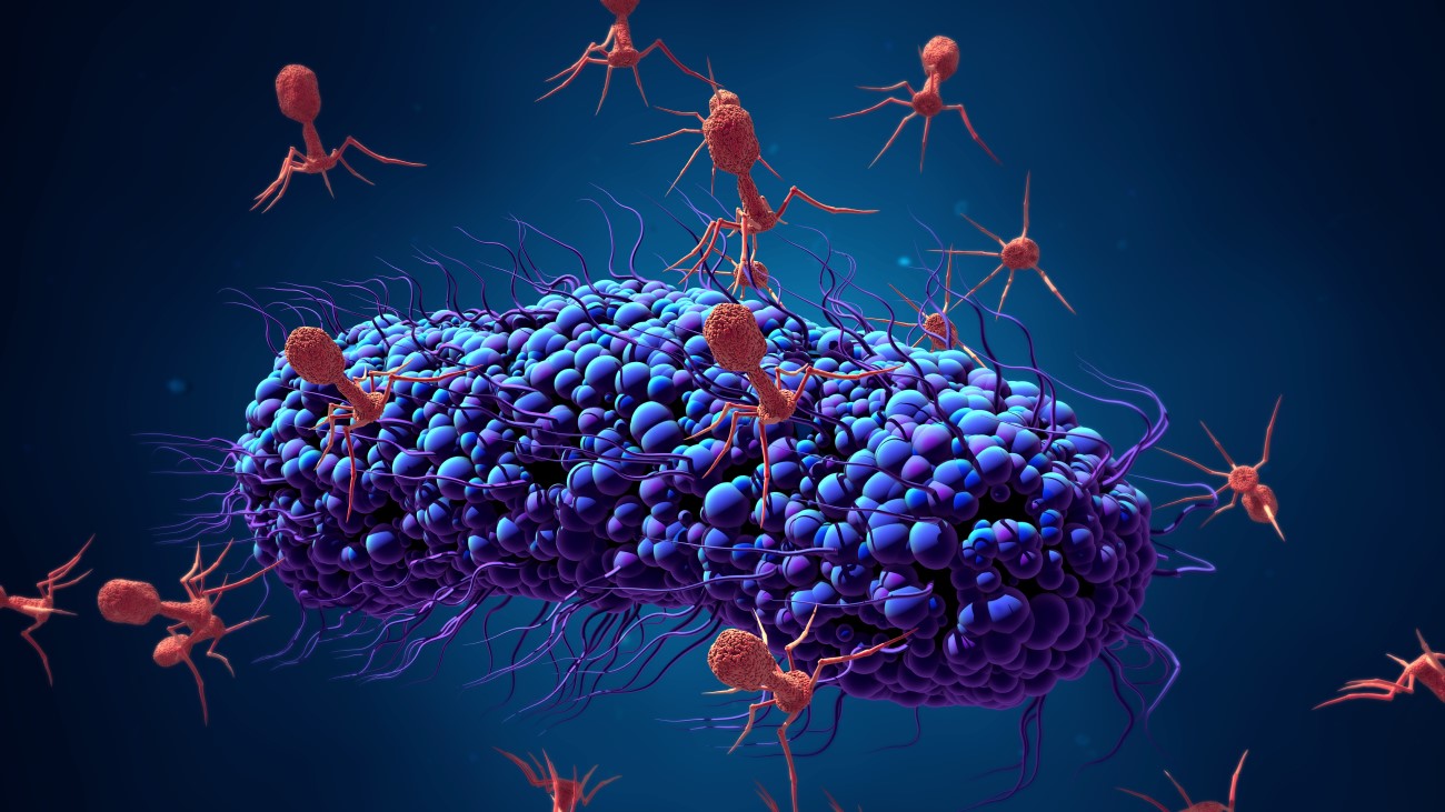
On Apr. 17, 2025, researchers from the University of of Pittsburgh announced they have produced the most detailed image to date of a bacteriophage, or phage — a kind of virus that kills bacteria — that has allowed them to see for the first time the structural makeup of the part of the virus that directly attaches to its target Mycobacterium cell. The work could enable new therapies that use bioengineered phages.
That’s important because of what a bacteriophage, or phage for short, does after it binds to a bacterial cell: it pierces a hole in the cell membrane and injects its own DNA, turning the bacteria into a phage factory.
With many bacteria growing increasingly resistant to the antibiotics we use to kill them, phages are in some cases the only option to fight off bacterial infections. They are, however, picky assassins: A particular kind of phage will typically attack just one strain of bacteria. The ability to engineer a phage to seek out and destroy a specific bacteria could be a medical game-changer.
The images are made dynamic thanks to work done by Raphael Park and his colleagues at Scripps Research, who used another kind of imaging, cryo-electron tomography, which images phages bound to the bacterial cell, highlighting the entire phage and where it attaches to the bacterial cell surface. Their high-definition images have allowed them to see structures that had previously only been resolved to a fuzzy grayscale that indicated the density of electrons.
“Now you can show every molecular component in this thing,” said researcher Krista Freeman. “And it’s just breathtaking.” The new images also reveal structural information that researchers can explore to better understand the point of contact between a phage and its bacterial target. The research was published in the journal Cell.
Tags:
Source: University of Pittsburgh
Credit: