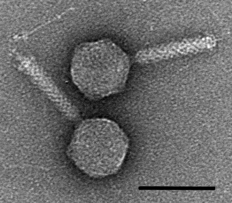
The electron microscope was used to characterize a bacteriophage virus that infects bacteria
In 1942, the first phage electron micrographs (EM) were published in 1940 in Germany and proved the particulate nature of bacteriophages. Phages and infected bacteria were first examined raw and unstained.
EM is heavily indebted to the brothers Ernst and Helmut Ruska. The former devoted himself to technical developments and the second concentrated on biological electron microscopy. In 1940, H. Ruska and Pfankuch and Kausche, all working in the Siemens & Halske laboratory, published two short papers submitted the same day to the same journal and showing the first pictures of bacteriophages in the world literature.
Tags:
Source: U.S. National Library of Medicine
Credit: Photo: Transmission electron microscopy of the V22 phage (species: Myoalterovirus PT11V22, morphotype: Myovirus, host: Alteromonas mediterranea PT11 ). The morphology of V22 is distinctly myoviral featuring. V22 has an icosahedral capsid (ø79 ± 5 nm) and a noncontracted tail length of 121 ± 11 nm with a fiberless baseplate complex (20 ± 3.7 nm height). Dimensions calculated as mean ± SD, n = 6. Bar, 100 nm. Courtesy: Rafael Gonzalez-Serrano, Matthew Dunne, Riccardo Rosselli, Ana-Belen Martin-Cuadrado, Virginie Grosboillot, Léa V. Zinsli, Juan J. Roda-Garcia, Martin J. Loessner, and Francisco Rodriguez-Valera
