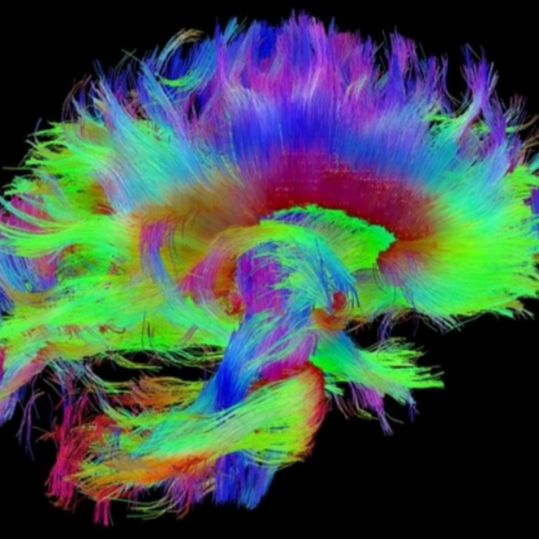
NIH study showed highly reproducible sex differences in aspects of human brain anatomy
On Jul. 20, 2020, a scientific analysis of more than 2,000 brain scans found evidence for highly reproducible sex differences in the volume of certain regions in the human brain. This pattern of sex-based differences in brain volume corresponds with patterns of sex-chromosome gene expression observed in postmortem samples from the brainメs cortex, suggesting that sex chromosomes may play a role in the development or maintenance of sex differences in brain anatomy. The study, led by researchers at the National Institute of Mental Health, was published in Proceedings of the National Academy of Sciences.
Researchers have long observed consistent sex-based differences in subcortical brain structures in mice. Some studies have suggested these anatomical differences are largely due to the effects of sex hormones, lending weight to a “gonad-centric” explanation for sex-based differences in brain development. However, more recent mouse studies have revealed consistent sex differences in cortical structures, as well, and gene-expression data suggest that sex chromosomes may play a role in shaping these anatomical sex differences. Although the mouse brain shares many similarities with the human brain, it is not clear whether these key findings in mice also apply to humans.
To explore the neurobiological basis of sex differences in the human brain, Raznahan, lead author Siyuan Liu, Ph.D., and colleagues first analyzed neuroimaging data collected as part of the Human Connectome Project (HCP). The data, obtained from 976 healthy adults between the ages of 22 and 35, revealed consistent sex differences in the volume of certain cortical structures. On average, females had relatively greater cortical volume in the medial and lateral prefrontal cortex, orbitofrontal cortex, superior temporal cortex, and lateral parietal cortex. Males, on average, had relatively greater cortical volume in ventral temporal regions and occipital regions, including the temporal pole, fusiform gyrus, and primary visual cortex.
Liu and colleagues then used two complementary approaches to determine whether these findings were reproducible. First, the researchers conducted 1,000 split-half comparisons by randomly splitting the HCP dataset in half and comparing results in the two halves. The results of these split-half comparisons indicated that the pattern of sex-based differences in cortical volume was highly stable. Second, the researchers quantified the reproducibility of the HCP findings in an unrelated neuroimaging dataset from the UK Biobank. Although the datasets had notable demographic and methodological differences, the researchers found that the overall pattern of sex-based differences in cortical volume was highly consistent.
Liu and co-authors then cross-referenced their anatomical findings with publicly available maps of gene-expression in the brain, which are based on 1,317 postmortem tissue samples from six human donors. The results indicated that the spatial pattern of sex differences in cortical volume was similar to the spatial pattern of sex-chromosome gene expression in the cortex. Specifically, regions of the cortex with relatively high expression of sex-chromosome genes tended to have greater cortical volume in males than females.
This correspondence with cortical expression of sex-chromosome genes is also consistent with findings from earlier mouse studies, suggesting that sex differences in brain anatomy may be due, at least in part, to genetic mechanisms that have been conserved throughout mammalian evolution. These findings suggest that sex differences in cortical volume may be influenced by genes located on the X and Y sex chromosomes.
The researchers also compared the anatomical findings with data from more than 11,000 functional neuroimaging studies. The results indicated spatial overlap between areas of the brain that showed sex-based differences in cortical volume in the HCP dataset and areas of the brain associated with face processing in functional neuroimaging studies.
Taken together, these findings shed light on the mechanisms that may contribute to sex-based differences in brain anatomy and point to genetic factors that may contribute to sex-based differences in brain disease and behavior. With these correlational findings as a roadmap, future research can more efficiently investigate the causes and consequences of sex differences in the human brain.
Tags:
Source: National Institutes of Health
Credit: